X-ray Spectroscopies

X-ray spectroscopies are techniques that use X-rays as a probe to investigate matter. The network offers various instruments that utilize X-ray tubes as sources. Specifically, the instruments can be divided into two categories: X-ray fluorescence and radioluminescence systems. In the case of X-ray fluorescence, the source excites the inner-shell electrons of atoms, and following relaxation processes, the electrons return to their ground state, releasing energy in the form of X-rays. The re-emitted X-rays will have an energy different from that of the incident X-rays, and this energy is characteristic of each element. The X-ray emission spectrum therefore allows for both the identification of the atoms present in the sample and their quantitative analysis. In radioluminescence, the emission of UV-visible light from a sample is studied under X-ray irradiation. This technique is essential for the characterization of scintillator materials, which form the basis of detectors for ionizing radiation. Radioluminescence is often coupled with thermoluminescence measurements. In this case, after irradiating the sample, the emission of thermally stimulated light is studied. This technique is particularly useful for characterizing defects in scintillator materials and is also applied in the dating of ceramic materials.

Our Instruments

The Network includes several instruments for X-ray fluorescence and a system for radioluminescence and thermoluminescence
X-ray fluorescence

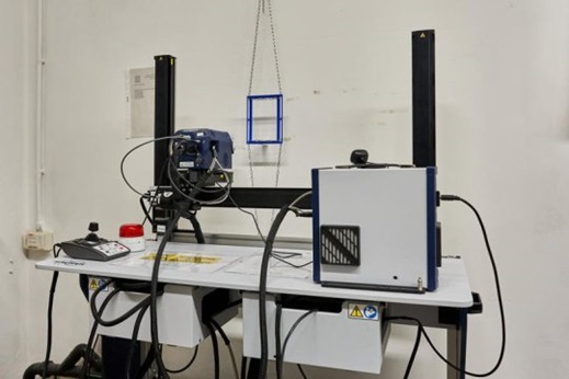
IRIS COMBINED X-RAY FLUORESCENCE & REFLECTANCE SPECTROSCOPY SCANNER SYSTEM

The IRIS Combined X-Ray Fluorescence & Reflectance Spectroscopy Scanner System is a mobile device that enables simultaneous scanning of a large area using both X-ray fluorescence spectroscopy (MA-XRF) and Vis-NIR reflectance spectroscopy. The X-ray fluorescence (MA-XRF) scanning technique allows for multi-element mapping of macro-areas, focusing on elements with an atomic number greater than 12 (Z>12). The reflectance Vis-NIR spectroscopy scan captures hyperspectral images across a wide spectral range, providing detailed information about the distribution and chemical nature of both inorganic and organic components. Data from both techniques are integrated for each point on the scanned surface, combining elemental chemical information with molecular data.
Technical specifications:
● halogen lamp: 360 – 2400 nm, nominal wattage: 20W);
● two detectors: 2048 channels, spectral range 400-1100 nm, 1.5 nm resolution;
512 channels, spectral range 1100-2500 nm, 8.9 nm resolution;
● X-ray tube with rhodium anode: window: Be 250 μm; 50 kV; 200 μA; collimators: 0.5 mm, 1 mm or 2 mm. Optional transmission filters;
● SSD detector: active area: 50 mm2, detector thickness: 450 μm, Mn-Kα resolution: 140 eV, dead layer thickness: 0.15 μm, window: Be 12.5 μm;
● XYZ motorised framework for making maps.
Available configurations:
● the system is designed for fast and non-invasive in situ scanning of the sample.
Applications:
● combined study of the distribution of atomic and molecular species in a sample;
● characterisation of inorganic and organic materials;
●analysis of painting techniques;
● diagnostic support for the restauration and conservation of artefacts.

Artax 200 XRF Bruker

The Artax 200 is a portable XRF instrument used for cultural heritage applications thanks to situ and non-invasive measurements. The system performs simultaneous multi-element analysis with atomic number greater than or equal to 13.
Technical specifications:
● source: Mo anode X-ray tube (10 – 50 kV, 5 µA min – 200 µA max);
● detector: Large Silicon Drift with 25 mm2 active area, window: 12 µm Be;
● resolution: less than 135 eV at MnKα with Peltier cooling.
Available configurations:
● “grazing-emission” measurements.
Applications:
● pigment analysis of paintings and frescoes and identification of possible restauration;
● development of new methodologies to optimise XRF data acquired with scanner instruments in order to obtain elemental distribution maps.

Radio and thermoluminescence

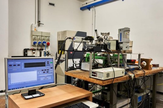
Home-made apparatus for radio and thermoluminescence measurements

This laboratory-developed instrumental apparatus is designed for wavelength- and temperature-resolved radioluminescence and thermoluminescence measurements. The instrument can measure light emitted under X-radiation (radioluminescence) at a constant temperature, or thermally stimulated emission (thermoluminescence).
Technical specifications:
● source: X-ray (10 – 40 kV);
● temperature range: 10-320 K.
Available configurations:
● measurements on solid and powder samples;
● for liquid samples, radioluminescence measurement is possible at room temperature or the solution can be deposited in metal crucibles for measurement.
Applications:
● characterisation and study of defects in luminescent materials through scintillation and dosimetry.

Contact us

If you believe these techniques could be useful for characterizing, updating, or designing your materials, please feel free to contact us