Raman Spectroscopy

Raman spectroscopy is a non-invasive, non-destructive technique used to investigate the chemical composition and molecular structure of samples. It is based on the inelastic scattering of monochromatic light by the sample. The frequencies of the scattered light depend on the material’s composition and structure, allowing for the identification and characterisation of the sample.
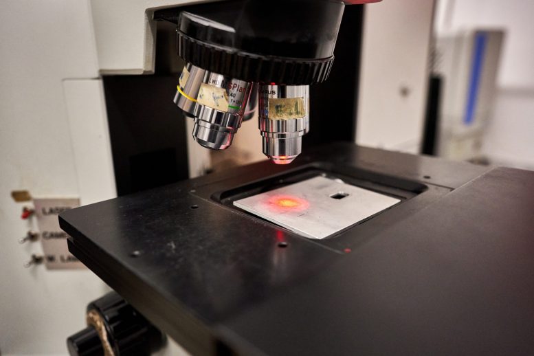
In a micro-Raman system, the Raman spectrometer is integrated with a microscope, which focuses the laser onto the sample. This enables the analysis of small regions (in the micrometer scale) or confocal analyses, where only the signal from a specific layer of the sample is studied. This feature allows for detailed analysis of layered or three-dimensional structures.
Raman spectroscopy provides valuable information about chemical bonds and three-dimensional structures. Specifically, it allows the study of the symmetry and orientation of anisotropic materials, the presence of tensile or compressive stresses, chemical composition, crystallinity, polymorphism, the presence of impurities, and can be used to estimate the homogeneity of a sample.
Additionally, Raman analysis does not require sample preparation, which is particularly important for in-situ applications, such as for studying artistic works (even under glass) or stone materials.
Raman spectroscopy is widely used in the study of materials in the solid state, whether crystalline or amorphous (semiconductors, polymers, coatings, composites, carbon nanotubes, graphene, ceramics, glass, gems…), but also for liquid samples and, if required even gaseous ones (e.g. gas inclusions in rocks).

Our instruments

The network includes a wide variety of Raman spectrophotometers coupled with microscopes (micro-Raman), equipped with different laser sources, sample holders, and objectives for magnification and analysis at various depths.
&nmsp;
Micro-Raman Labram Dilor Jobin Yvon

The Labram Dilor Raman micro-spectrometer operates in a back-scattering configuration and is confocally coupled to a microscope. The microscope (BX40, Olympus) allows for the selective analysis of samples at the micrometer scale. Its various objectives enable the selection of different sample areas and the analysis of different depths, such as in multi-layer samples, thanks to their varying numerical apertures.
Technical specifications:
● laser sources: Ar+ at 488 nm (maximum power 50 mW), He-Ne at 633 nm (maximum power 20mW);
● sample holder: motorised xy stage characterised by a lateral resolution of approximately 1 μm and a vertical resolution of up to 2 μm;
● microscope objectives: 10X (0.25NA), 20X (0.4 NA), 50X (0.7 NA), 100 X (0.9 NA), 50X-LWD (long work distance, 0.75 NA);
● spectrometer resolution: 1 cm-1.
Available configurations:
● variable temperature measurements (4 K ÷ 500 K) and micro-luminescence measurements excited at 488 nm and 633 nm.
Applications:
● study of materials for photonics and optoelectronics applications;
● identification of crystalline phases in gemological stones;
● polymers identification;
● characterisation of carbon-based materials (carbon black, graphite, graphene, nanotubes, amorphous carbon, diamond);
● determination of inorganic pigments;
● in situ and in operando study of fuel cells and batteries.

Micro-Raman T64000 Jobin Yvon

The micro-Raman T64000 is a modular instrument with a flexible configuration. The system includes a microscope that illuminates the sample and collects the inelastically scattered light, which is then analysed by a spectrograph coupled with a double pre-monochromator.
Technical specifications:
● spectral range: UV-Vis;
● excitation sources: multiple options available;
● diffraction-limited spatial resolution (< 1 µm);
● motorised XY stage with 0.1 µm steps.
Available configurations:
● confocal mode;
● polarised light analysis.
Applications:
● characterisation of materials for microelectronics and optoelectronics.

Micro-Raman LabRAM Evolution Horiba Jobin Yvon

The LabRAM HR Evolution Raman spectrometer is a system coupled with an Olympus BXFM petrographic microscope for observations in both transmitted and reflected light, connected to a 5 MP camera
The presence of a motorized sample stage and a confocal system enables the acquisition of both two- and three-dimensional profiles and maps.
Technical specifications:
● source: 532 nm green Nd laser (power: 300 mW);
● microscope objectives: 5X, 10X, 50X LWD and 100X;
● sample holder: motorised.
Available configurations:
● the sample stage is compatible with a LINKAM THMS600 heating/freezing table for studying fluid inclusions.
Applications:
● analysis of minerals and rocks: thin/double glazed sections;
● analysis of micro- and nano-plastics.

Micro-Raman InVia Renishaw

The inVia Reflex spectrometer is a system composed of a high-performance Raman spectrometer coupled with a Leica DM2500 polarized light research microscope.
Technical specifications:
● laser sources: solid-state at 532 nm (maximum power 50 mW) and 785 nm;
● sample holder: motorised stage x-y;
● microscope objectives: 5x, 20x, 50x, 50×LWD, 100x;
● high resolution camera: 5.1 MP;
● software: Latest WIRE 5.5 (2024).
Available configurations:
●The HSES motorised base, developed specifically for StreamLine rapid mapping, ensures extreme accuracy in sample positioning. New acceleration algorithms enable fast, accurate, and repeatable movements of the stage across its entire range.
Applications:
● Mineralogy of sediments:
characterisation of the mineralogical composition of sediments transported by rivers, deserts and beaches;
characterisation of the mineralogical composition of marine sediments in turbiditic and contouritic deposits;
creation of databases for mineral recognition.
● Study of climate change:
mineralogical analysis of dust in ice cores from Alpine, Arctic and Antarctic glaciers down to 1 micron.
● Industrial applications:
study of inclusions and impurities in glass of industrially produced bottles.
● Archaeology:
characterisation of the mineralogical composition of ceramics and lithic artefacts.
● Plastics and textile fibres:
identification of microplastics and fibres in sediments, seawater and the atmosphere;
creation of databases for recognition of plastics and fibres.
● Analysis of tumour cells.

Micro-Raman Qontor Renishaw

The Qontor spectrometer is a system composed of a high-performance Raman spectrometer and a Leica DM2500 polarised light research microscope.
Technical specifications:
● laser sources: at 630 nm and at 532 nm;
● sample holder: motorised stage x-y;
● autofocus in z-axis;
● microscope objectives: 2.5x, 5x, 10x, 50×LWD, 100x;
Available configurations:
●The HSES motorized base, developed specifically for StreamLine rapid mapping, ensures exceptional accuracy in sample positioning. New acceleration algorithms enable fast, accurate, and repeatable stage movements of the stage across its entire range.
Applications:
characterisation of the metamict state of zircons for applications in U-Pb dating;
● Raman maps (2D and 3D) for high-resolution chemical imaging with LiveTrack real-time focus;
● Raman maps to identify the coordinates of specific minerals of interest for Geochronology (zircon, apatite, monazite);
● varietal studies of mineral groups using internal standards and databases (Feldspars, Carbonates, Phosphates, Amphiboles, Epidotes, Garnets, Pyroxenes).

i-Raman Plus BW Tec

The i-Raman Plus BW Tec is a portable instrument, thanks to its compact size and low power consumption.
Technical specifications:
● source: 785 nm laser (maximum power 418 mW);
● spectral range: 200 – 3000 cm-1;
● resolution: below 5 cm-1;
● probe: optical fibre.
Available configurations:
● adjustable power output of the source;
● the optical fibre probe can be used with a probe holder for analysing liquid samples or with an XYZ stage for analysing samples on the plane or with a video microscope.
Applications:
●  identification of pigments or degradation products of artefacts;
●  evaluation of the state of degradation or corrosion of surfaces;
● study of ageing mechanisms and identification of production technologies or surface treatments.

Contact us

If you believe these techniques could be useful for characterizing, updating, or designing your materials, please feel free to contact us